The origins of cell- and microbiology: Hooke and Leeuwenhoek
by Anders Skovly, 2025
Robert Hooke and the discovery of “cells”
The history of cell- and microbiology is tied up with the development of the early microscopes. One of the most famous men in early microscopy is Robert Hooke, who in 1665 published the book Micrographia. In this book Hooke describes various objects as seen under his microscope, and supplemented his descriptions with some nicely drawn illustrations.
From the perspective of cell biology there is one particular observation in Micrographia that is of special interest. This is the observation on the microscopic structure of cork, the material best known for its use in wine bottle stoppers. Cork is one of the layers in the bark of trees. The layer of cork is usually quite thin, but in the bark of the cork oak the layer of cork can be many centimeters thick, and it is from this bark that cork products are made. Here is a quote from Micrographia, from Hooke's observation on cork:
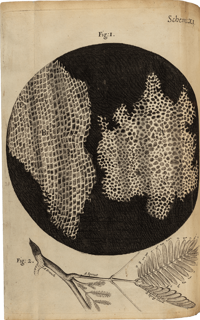
Observ. XVIII. Of the Schematisme or Texture of Cork, and of the Cells and Pores of some other such frothy Bodies.
I took a good clear piece of Cork, and with a Pen-knife sharpen’d as keen as a Razor, I cut a piece of it off, and thereby left the surface of it exceeding smooth, then examining it very diligently with a Microscope, me thought I could perceive it to appear a little porous; but I could not so plainly distinguish them, as to be sure that they were pores, much less what Figure they were of: But judging from the lightness and yielding quality of the Cork, that certainly the texture could not be so curious, but that possibly, if I could use some further diligence, I might find it to be discernable with a Microscope, I with the same sharp Pen-knife, cut off from the former smooth surface an exceeding thin piece of it, and placing it on a black object Plate, because it was it self a white body, and casting the light on it with a deep plano convex Glass, I could exceeding plainly perceive it to be all perforated and porous, much like a Honey-comb, but that the pores of it were not regular; yet it was not unlike a Honey-comb in these particulars.
First, in that it had a very little solid substance, in comparison of the empty cavity that was contain’d between, as does more manifestly appear by the Figure A and B of the XI. Scheme, for the Interstitia, or walls (as I may so call them) or partitions of those pores were neer as thin in proportion to their pores, as those thin films of Wax in a Honey-comb (which enclose and constitute the sexangular cells) are to theirs.
Source: Robert Hooke's Micrographia (1665).
Next, in that these pores, or cells, were not very deep, but consisted of a great many little Boxes, separated out of one continued long pore, by certain Diaphragms, as is visible by the Figure B, which represents a sight of those pores split the long-ways.
(Micrographia is freely available online at gutenberg.org in the form of html text. The book is also available at royalsociety.org in the form of a digitalized version of the original 1665 book.)
Note how Hooke describes the “pores” of cork as resembling the cells of a honeycomb. When we in the modern biology speak of “cells”, the above paragraphs is where the word “cell” supposedly originated. (I want to mention that what we in modern biology refer to as a cell, and what Hooke was referring to as a cell, are not exactly the same things. I will clarify this in a later article.)
Hooke also looks at other plant sections, including the pith of an Elder tree, and again he sees these "long pores" separated into "boxes" by "diaphragms". In many plant sections he finds the "pores" filled with "juices", and he considers that the diaphragms may function as valves to ensure one-way flow of the juice through the pores. He tries to test this by blowing into the wood:
But though I could not with my Microscope, nor with my breath, nor any other way I have yet try’d, discover a passage out of one of those cavities into another, yet I cannot thence conclude, that therefore there are none such, by which the Succus nutritius, or appropriate juices of Vegetables, may pass through them; for, in several of those Vegetables, whil’st green, I have with my Microscope, plainly enough discover’d these Cells or Pores fill’d with juices, and by degrees sweating them out; as I have also observed in green Wood all those long Microscopical pores which appear in Charcoal perfectly empty of any thing but Air.
Now, though I have with great diligence endeavoured to find whether there be any such thing in those Microscopical pores of Wood or Piths, as the Valves in the heart, veins, and other passages of Animals, that open and give passage to the contain’d fluid juices one way, and shut themselves, and impede the passage of such liquors back again, yet have I not hitherto been able to say any thing positive in it; though, me thinks, it seems very probable, that Nature has in these passages, as well as in those of Animal bodies, very many appropriated Instruments and contrivances, whereby to bring her designs and end to pass, which ’tis not improbable, but that some diligent Observer, if help’d with better Microscopes, may in time detect.
(It is now known that the diaphragms in the long pores do not function as valves. I'm no plant biologist, so I won't give further comment on their function.)
The microscope used by Hooke is described in Micrographia’s preface and is illustrated in Scheme I. It had two lenses: one at the “bottom” (near the object to be looked at), and one at the “top” (where you put your eye), similar to a modern compound microscope (that is, a microscope with multiple lenses). Hooke's microscope had relatively low magnification by today’s standards, and I don’t think one could have seen bacteria with it.
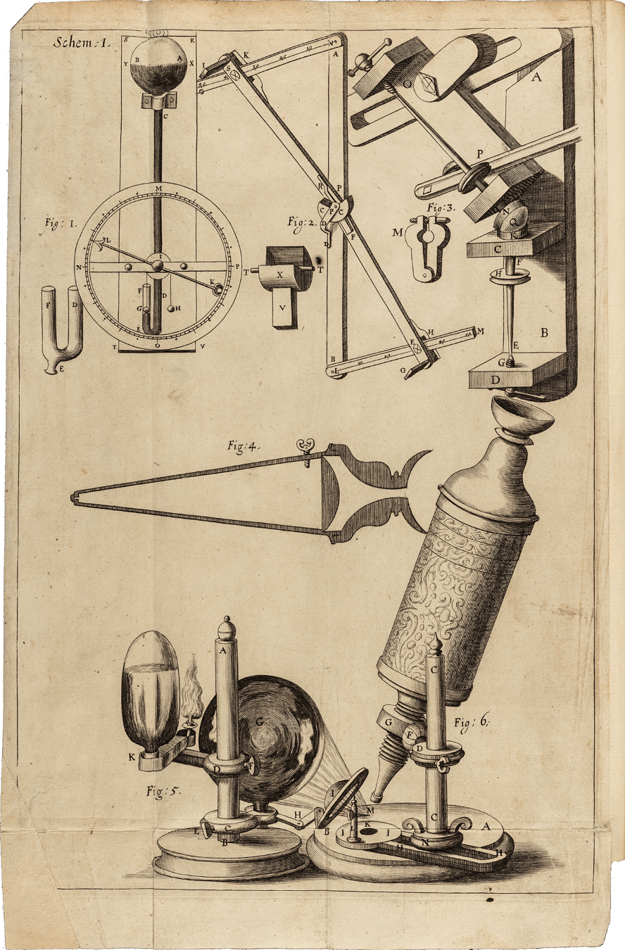
(the upper part of the figure is not related to the microscope).
Source: Robert Hooke's Micrographia (1665).
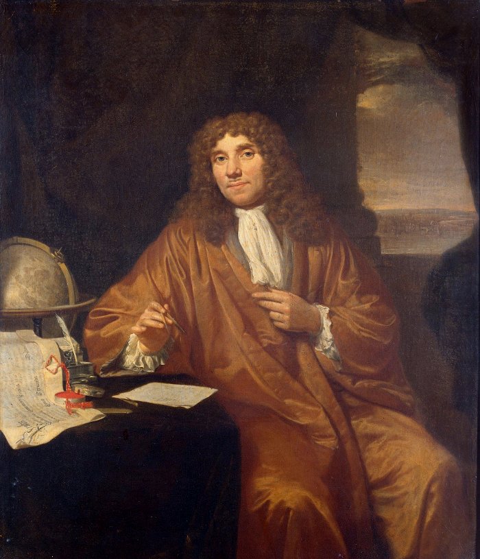
Antony van Leeuwenhoek and the discovery of “animalcules”
We now move on from Hooke to another character, namely Antony van Leeuwenhoek. Unlike Hooke, Leeuwenhoek did not communicate his observations in a book, but through a series of letters sent to the Royal Society of London and to others. His original texts were all written in Dutch, so here we will be dealing with english translations.
One of the best sources of information on Leeuwenhoek is the book Antony van Leeuwenhoek and his Little animals, written by the bacteriologist and protozoologist Clifford Dobell and published in 1932. This book contains a biography of Leeuwenhoek, as well as many translated segments of Leeuwenhoek’s letters, accompanied by Dobell’s comments to the letters. It is freely available on biodiversitylibrary.org.
Let’s look at the letter in which Leeuwenhoek described his first observation of microscopic organisms. This is letter nr. 11 [6], dated 7th September 1674, translated by Dobell:
About two hours distants from this Town [Delft, in the Netherlands] there lies an inland lake, called the Berkelse Mere, whose bottom in many places is very marshy, or boggy. Its water is in winter very clear, but at the beginning or in the middle of summer it becomes whitish, and there are then little green clouds floating through it; which, according to the saying of the country folk dwelling thereabout, is caused by the dew, which happens to fall at that time, and which they call honey-dew. This water is abounding in fish, which is very good and savoury.
Passing just lately over this lake, at a time when the wind blew pretty hard, and seeing the water as above described, I took up a little of it in a glass phial; and examining this water next day, I found floating therein divers earthy particles, and some green streaks, spirally wound serpent-wise, and orderly arranged, after the manner of the copper or tin worms, which distillers use to cool their liquors as they distil over. The whole circumference of each of these streaks was about the thickness of a hair of one’s head. Other particles had but the beginning of the foresaid streak; but all consisted of very small green globules joined together: and there were very many small green globules as well.
Among these there were, besides, very many little animalcules [very small animals], whereof some were roundish, while others, a bit bigger, consisted of an oval. On these last I saw two little legs near the head, and two little fins at the hindmost end of the body. Others were somewhat longer than an oval, and these were very slow a-moving, and few in number. These animalcules had divers colours, some being whitish and transparent; others with green and very glittering little scales; others again were green in the middle, and before and behind white; others yet were ashen grey. And the motion of most of these animalcules in the water was so swift, and so various, upwards, downwards, and round about, that ’twas wonderful to see: and I judge that some of these little creatures were above a thousand times smaller than the smallest ones I have ever yet seen, upon the rind of cheese, in wheaten flour, mould, and the like.
Dobell comments that the “green streaks” were “The common green alga Spirogyra: the earliest recorded observations on this organism.” He further says that the animalcules with “two little legs near the head, and two little fins at the hindmost end of the body” were probably Rotifiers, and that the animalcules that were “somewhat longer than an oval, and these were very slow a-moving” were probably Ciliates. Finally, he remarks that when Leeuwenhoek says “some of these little creatures were above a thousand times smaller than the smallest ones I have ever yet seen”, Leeuwenhoek means a thousand times smaller in volume, not in length or width. Therefore, Leeuwenhoek judged these new organisms to be over ten times smaller, in terms of length/width, than anything he’d seen before.
In the article “The riddle of the green streaks”, Wim van Egmond writes that the green streaks were probably not the alga Spirogyra, but rather the cyanobacteria Dolichospermum. His article features microscope-photographs of both Spirogyra and Dolichospermum.
Rotifers, aka wheel animals, can be seen in this video. (The rotifiers don’t actually have wheels, they just have something that looks like wheels when seen with a microscope.)
Here is another excerpt, this one from Leeuwenhoek’s letter nr. 26 [18], dated 9th October 1676, translated by Dobell. It describes a curious organism discovered in rainwater collected in a pot:
In the year 1675, about half-way through September (being busy with studying air, when I had much compressed it by means of water), I discovered living creatures in rain, which had stood but a few days in a new tub, that was painted blue within. This observation provoked me to investigate this water more narrowly; and especially because these little animals were, to my eye, more than ten thousand times smaller [in volume] than the animalcule which Swammerdam has portrayed, and called by the name of Water-flea, or Water-louse, which you can see alive and moving in water with the bare eye.
Of the first sort that I discovered in the said water, I saw, after divers observations, that the bodies consisted of 5, 6, 7, or 8 very clear globules, but without being able to discern any membrane or skin that held these globules together, or in which they were inclosed. When these animalcules bestirred ’emselves, they sometimes stuck out two little horns, which were continually moved, after the fashion of a horse’s ears. The part between these little horns was flat, their body else being roundish, save only that it ran somewhat to a point at the hind end; at which pointed end it had a tail, near four times as long as the whole body, and looking as thick, when viewed through my microscope, as a spider’s web. At the end of this tail there was a pellet, of the bigness of one of the globules of the body; and this tail I could not perceive to be used by them for their movements in very clear water.
These little animals were the most wretched creatures that I have ever seen; for when, with the pellet, they did but hit on any particles or little filaments (of which there are many in water, especially if it hath but stood some days), they stuck intangled in them; and then pulled their body out into an oval, and did struggle, by strongly stretching themselves, to get their tail loose; whereby their whole body then sprang back towards the pellet of the tail, and their tails then coiled up serpentwise, after the fashion of a copper or iron wire that, having been wound close about a round stick, and then taken off, kept all its windings. This motion, of stretching out and pulling together the tail, continued; and I have seen several hundred animalcules, caught fast by one another in a few filaments, lying within the compass of a coarse grain of sand.
Dobell comments that the organism here described is Vorticella, and that Leeuwenhoek later came to believe that the contraction of the Vorticella’s tail allows the Vorticella to pull water towards itself, so as to get food from the water. Unlike the Rotifer, the Vorticella is not classified as an animal, but is classified as belonging to a group of organisms called the “protists”. A video of Vorticella can be seen here.
(The original english translation of letter nr. 26 [18], published in the Philosophical Transactions of the Royal Society in 1677, is freely available at royalsocietypublishing.org. It’s not that easy to read it, as the old letter “s” looks like a letter “f”. This letter is considered to be the first time bacteria were described (I’m not going to quote that part of the letter here). However, if Leeuwenhoek’s “green streaks” described previously are in fact the cyanobacteria Dolichospermum, then that would be the first description of a bacteria.
Here’s a third excerpt, this one from letter 76 [39], dated 17th September 1683, and also translated by Dobell. It describes the various forms of organisms found in the human mouth:
... Yet notwithstanding, my teeth are not so cleaned thereby, but what there sticketh or groweth between some of my front ones and my grinders (whenever I inspected them with a magnifying mirror), a little white matter, which is as thick as if ’twere batter. On examining this, I judged (albeit I could discern nought a-moving in it) that there yet were living animalcules therein. I have therefore mixed it, at divers times, with clean rain-water (in which there were no animalcules), and also with spittle, that I took out of my mouth, after ridding it of air-bubbles (lest the bubbles should make any motion in the spittle): and I then most always saw, with great wonder, that in the said matter there were many very little living animalcules, very prettily a-moving.
The biggest sort had the shape of Fig. A: these had a very strong and swift motion, and shot through the water (or spittle) like a pike does through the water. These were most always few in number.
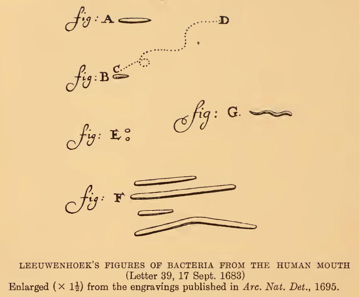
The second sort had the shape of Fig. B. These ofttimes spun round like a top, and every now and then took a course like that shown between C and D: and these were far more in number.
To the third sort I could assign no figure: for at times they seemed to be oblong, while anon they looked perfectly round. These were so small that I could see them no bigger than Fig. E: yet therewithal they went ahead so nimbly, and hovered so together, that you might imagine them to be a big swarm of gnats or flies, flying in and out among one another. These last seemed to me e’en as if there were, in my judgement, several thousand of ’em in an amount of water or spittle (mixed with the matter aforesaid) no bigger than a sand-grain; albeit there were quite nine parts of water, or spittle, to one part of the matter that I took from betwixt my front teeth, or my grinders.
Furthermore, the most part of this matter consisted of a huge number of little streaks, some greatly differing from others in their length, but of one and the same thickness withal; one being bent crooked, another straight, like Fig. F, and which lay disorderly ravelled together. And because I had formerly seen, in water, live animalcules that had the same figure, I did make every endeavour to see if there was any life in them; but I could make out not the least motion, that looked like anything alive, in any of ’em.
[...]
While I was talking to an old man (who leads a sober life, and never drinks brandy or tobacco, and very seldom any wine), my eye fell upon his teeth, which were all coated over; so I asked him when he had last cleaned his mouth? And I got for answer that he’d never washed his mouth in all his life. So I took some spittle out of his mouth and examined it; but I could find in it nought but what I had found in my own and other people’s.
I also took some of the matter that was lodged between and against his teeth, and mixing it with his own spit, and also with fair water (in which there were no animalcules), I found an unbelievably great company of living animalcules, a-swimming more nimbly than any I had ever seen up to this time. The biggest sort (whereof there were a great plenty) bent their body into curves in going forwards, as in Fig. G. Moreover, the other animalcules were in such enormous numbers, that all the water (notwithstanding only a very little of the matter taken from between the teeth was mingled with it) seemed to be alive. The long particles too, as before described, were also in great plenty.
Dobell remarks that all the organisms here described are bacteria, and that the mouth is the only place where so many different shapes of bacteria can be found together.
While Clifford Dobell’s book is one source of translations of Leeuwenhoek’s letters, his book deals chiefly with the letters concerning the microscopic organisms we now call bacteria and protozoa. A Dutch organization has been working on english translations of all of Leeuwenhoek’s scientific letters. The first part of this work was published in 1939 under the name “The Collected Letters of Antony van Leeuwenhoek, Volume 1”. At the time of writing this article, 17 volumes have been published, with the last few volumes scheduled to be published soon. The first 15 volumes are freely available on the website of the Digitale Bibliotheek voor de Nederlandse Letteren.
Based on the little I’ve read of these Collected Letters I want to show one last excerpt from Leeuwenhoek’s writings. This is from Volume 3, letter 67 [35], published 3rd March 1682:
After inspecting with astonishment the numerous flesh-muscles of an ox-tail, I intended to observe the tail of a ray, but while cutting the tail I saw the blood gushing from it. Observing this blood it struck me that the parts of the blood that are globules in human beings and mammals and colour the blood red, are here all of them flat, oval particles, thickish, floating in a crystalline water. Where these oval particles lay single they had no colour, but when 3 or 4 lay on top of each other, they began to show a red colour. Thus I came to observe the blood of a cod and of a salmon, which I also found to consist of hardly anything but oval figures [...] represented in Fig. 5.
Source: dbnl.org
As we see here, Leeuwenhoek also used his microscopes to study the structure of animals, and he found that the blood of both fishes and humans consists predominantly of red blood cells (although he didn’t use the word “cell” for what he saw: Leeuwenhoek didn’t have the concept of cells, as this concept was developed in the 1800s).
Now we reach the final part of this text, where we consider the microscopes built and used by Leeuwenhoek. These look nothing like today’s typical idea of a microscope. Leeuwenhoek’s design was that of two metal plates sandwiching a tiny lens, with a screw mechanism for positioning and focusing a sample object in front of the lens. The other side of the microscope would then be held up to the eye, so that one can see through the lens onto the sample object. Pictures of eleven Leeuwenhoek microscopes are available on leeuwenhoek.net.
Video clips filmed through one of Leeuwenhoek’s microscopes showing bacteria, alga, Vorticella and Rotifier can be seen on visualizingtheunknown.com. And this video by the BBC demonstrates how Leeuwenhoek's microscopes were used.
Zuilen 1981 inspected eight of Leeuwenhoek’s microscopes and found that his “best” microscope had a magnification of 266x, while the others had magnifications ranging from 69x to 167x. Zuilen states that Leeuwenhoek produced over 500 microscopes, most of which have now been lost. It is therefore quite possible that Leeuwenhoek had managed to make a few microscopes with even higher magnifications than 266x.
Robert Hooke’s Micrographia describes a microscope similar to those that Leeuwenhoek would make years after Micrographia’s publication. Hooke even pointed out that a microscope with a single lens was optically superior to larger compound microscopes such as the one he used himself. The appeal of the compound microscope was that it was more convenient to work with. Here quoting from the preface of Micrographia:
... if one of these [lenses] be fixt with a little soft Wax against a small needle hole, prick’d through a thin Plate of Brass, Lead, Pewter, or any other Metal, and an Object, plac’d very near, be look’d at through it, it will both magnifie and make some Objects more distinct then any of the great Microscopes. But because these, though exceeding easily made, are yet very troublesome to be us’d, because of their smalness, and the nearness of the Object; therefore to prevent both these, and yet have only two Refractions, I provided me a Tube of Brass, shap’d much like that in the fourth Figure of the first Scheme [...]
It would not be until sometime in the 1800’s that the optics of compound microscopes (with multiple lenses) began to outperform the optics of the single-lens microscopes.
References
Zuilen
(1981): The microscopes of Antoni van Leeuwenhoek. Paid article